


Bone Development & Growth
The terms osteogenesis and ossification are often used synonymously to indicate the process of bone formation. Parts of the skeleton form during the first few weeks after conception. By the end of the eighth week after conception, the skeletal pattern is formed in cartilage and connective tissue membranes and ossification begins.
Bone development continues throughout adulthood. Even after adult stature is attained, bone development continues for repair of fractures and for remodeling to meet changing lifestyles. Osteoblasts, osteocytes and osteoclasts are the three cell types involved in the development, growth and remodeling of bones. Osteoblasts are bone-forming cells, osteocytes are mature bone cells and osteoclasts break down and reabsorb bone.
There are two types of ossification: intramembranous and endochondral.
Intramembranous
Intramembranous ossification involves the replacement of sheet-like connective tissue membranes with bony tissue. Bones formed in this manner are called intramembranous bones. They include certain flat bones of the skull and some of the irregular bones. The future bones are first formed as connective tissue membranes. Osteoblasts migrate to the membranes and deposit bony matrix around themselves. When the osteoblasts are surrounded by matrix they are called osteocytes.
Endochondral Ossification
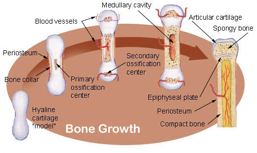
Endochondral ossification involves the replacement of hyaline cartilage with bony tissue. Most of the bones of the skeleton are formed in this manner. These bones are called endochondral bones. In this process, the future bones are first formed as hyaline cartilage models. During the third month after conception, the perichondrium that surrounds the hyaline cartilage "models" becomes infiltrated with blood vessels and osteoblasts and changes into a periosteum. The osteoblasts form a collar of compact bone around the diaphysis. At the same time, the cartilage in the center of the diaphysis begins to disintegrate. Osteoblasts penetrate the disintegrating cartilage and replace it with spongy bone. This forms a primary ossification center. Ossification continues from this center toward the ends of the bones. After spongy bone is formed in the diaphysis, osteoclasts break down the newly formed bone to open up the medullary cavity.
The cartilage in the epiphyses continues to grow so the developing bone increases in length. Later, usually after birth, secondary ossification centers form in the epiphyses. Ossification in the epiphyses is similar to that in the diaphysis except that the spongy bone is retained instead of being broken down to form a medullary cavity. When secondary ossification is complete, the hyaline cartilage is totally replaced by bone except in two areas. A region of hyaline cartilage remains over the surface of the epiphysis as the articular cartilage and another area of cartilage remains between the epiphysis and diaphysis. This is the epiphyseal plate or growth region.
Bone Growth
Bones grow in length at the epiphyseal plate by a process that is similar to endochondral ossification. The cartilage in the region of the epiphyseal plate next to the epiphysis continues to grow by mitosis. The chondrocytes, in the region next to the diaphysis, age and degenerate. Osteoblasts move in and ossify the matrix to form bone. This process continues throughout childhood and the adolescent years until the cartilage growth slows and finally stops. When cartilage growth ceases, usually in the early twenties, the epiphyseal plate completely ossifies so that only a thin epiphyseal line remains and the bones can no longer grow in length. Bone growth is under the influence of growth hormone from the anterior pituitary gland and sex hormones from the ovaries and testes.
Even though bones stop growing in length in early adulthood, they can continue to increase in thickness or diameter throughout life in response to stress from increased muscle activity or to weight. The increase in diameter is called appositional growth. Osteoblasts in the periosteum form compact bone around the external bone surface. At the same time, osteoclasts in the endosteum break down bone on the internal bone surface, around the medullary cavity. These two processes together increase the diameter of the bone and, at the same time, keep the bone from becoming excessively heavy and bulky.
Suggested Citation
SEER Training Modules: Bone Development & Growth. U.S. National Institutes of Health, National Cancer Institute. Cited 10 July 2026. Available from: https://training.seer.cancer.gov.