


Salpingo-Ovarian & Peritoneal Functional Anatomy
The ovaries are paired, oval-shaped organs measuring approximately 2-4 cm in diameter that lie on the posterior wall of the pelvis lateral to the uterus. They are supported by the suspensory ligaments, the ovarian ligament, and the broad ligament. The ovaries produce and expell ova (eggs) and function as glands that produce reproductive hormones.
The fallopian tubes are a pair of slender ducts, approximately 10 cm long with fingerlike projections adjacent to the ovary (fimbriae) through which ova pass from the ovaries to the uterus in the female reproductive system [4].
In females, the peritoneum, which includes the omentum and pelvic and abdominal viscera, lines the anterior (front) and posterior (back) surfaces of the uterus. It is more commonly a site of metastasis than of primary cancer [16].
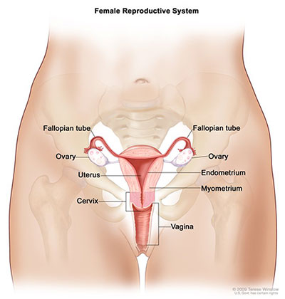
Anatomy of the female reproductive system.
Source: Terese Winslow (Illustrator), National Cancer Institute.
Anatomical Structures Related to the Ovaries
- Ovary
- Medial surface
- Lateral surface
- Free border
- Mesovarial margin
- Tubal extremity
- Uterine extremity
- Oviduct (fallopian tube)
- Opening of fallopian tube
- Infundibulum of fallopian tube
- Fimbriae of fallopian tube
- Ovarian fimbria
- Ampulla of fallopian tube
- Isthmus of fallopian tube
- Uterine part of fallopian tube
- Uterine opening of fallopian tube
Updated: June 8, 2018
Suggested Citation
SEER Training Modules: Salpingo-Ovarian & Peritoneal Functional Anatomy. U.S. National Institutes of Health, National Cancer Institute. Cited 09 June 2026. Available from: https://training.seer.cancer.gov.